New publication by Gabriels et al. in The Journal of Nuclear Medicine

Our latest publication by Ruben Gabriels entitled ‘Detection of early esophageal neoplastic Barrett lesions with quantified fluorescence molecular endoscopy using cetuximab-800CW’ can be found here.
Abstract
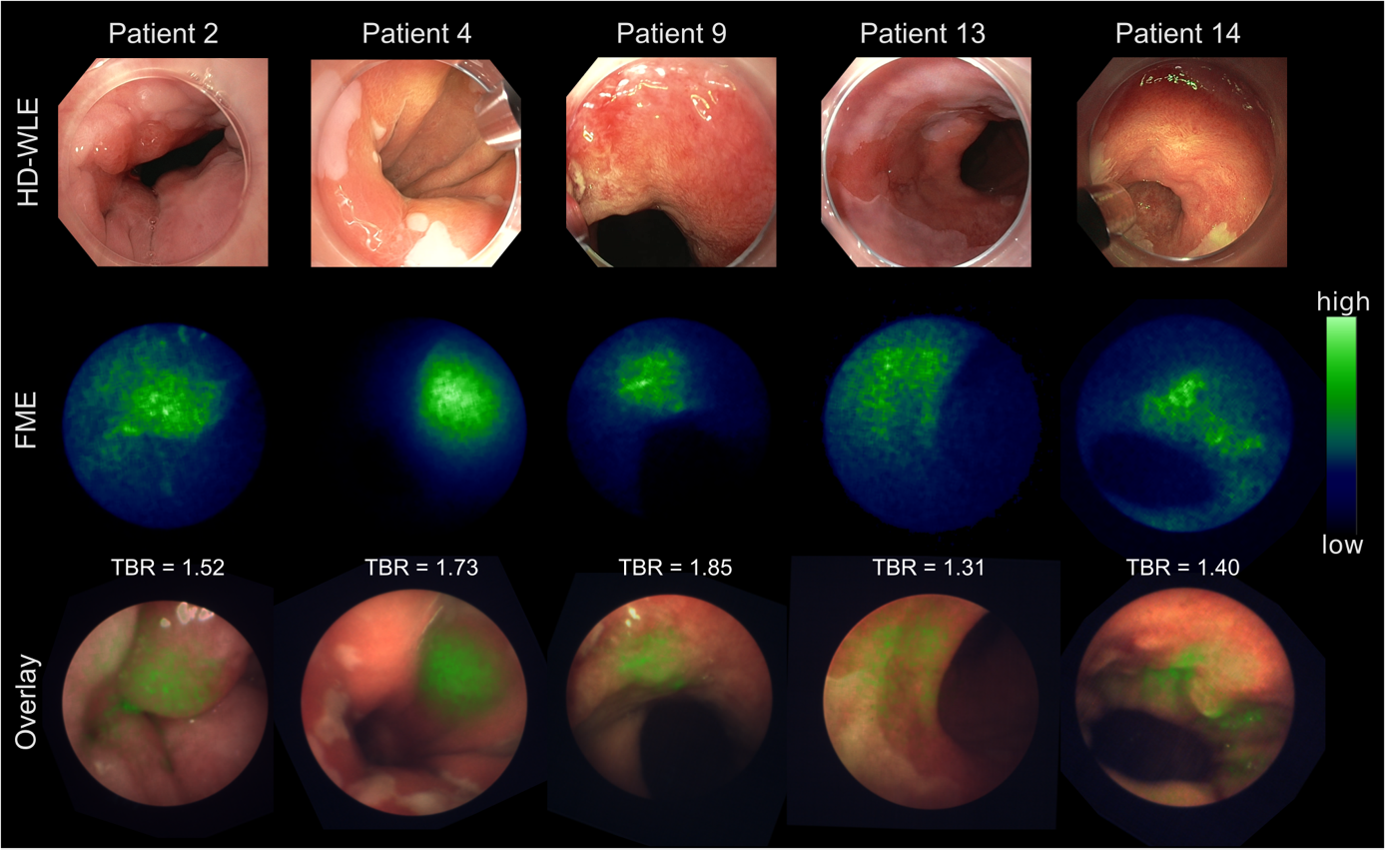
Esophageal adenocarcinoma (EAC) causes 6 % of cancer-related deaths worldwide. Near-infrared fluorescence molecular endoscopy (NIR-FME) uses a tracer that targets overexpressed proteins. In this study we aim to investigate the feasibility of an epidermal growth factor receptor (EGFR) targeted tracer, cetuximab-800CW, to improve detection of early-stage EAC. Methods: We validated EGFR expression in 73 esophageal tissue sections. Subsequently, we topically administered cetuximab-800CW and performed high-definition white-light endoscopy (HD-WLE), narrow band imaging (NBI) and NIR-FME in fifteen patients with Barrett’s esophagus (BE). Intrinsic fluorescence values were quantified using multi-diameter single fiber reflectance (MDSFR) and single-fiber fluorescence (SFF) spectroscopy. Back-table imaging, histopathological examination and EGFR immunohistochemistry on biopsies collected during NIR-FME procedures were performed and compared to in vivo imaging results. Results: Immunohistochemical pre-analysis showed high EGFR expression in 67% of dysplastic tissue sections. NIR-FME visualized all 12 HD-WLE visible lesions and 5 HD-WLE invisible dysplastic lesions, with increased fluorescence signal in visible dysplastic BE lesions compared to non-dysplastic BE as shown by MDSFR/SFF, reflecting a target-to-background ratio (TBR) of 1.5. Invisible dysplastic lesions also showed increased fluorescence with a TBR of 1.67. Immunohistochemistry analysis showed EGFR overexpression in 16 out of 17 (94%) dysplastic BE lesions, which all showed fluorescence signal. Conclusion: This study has shown that NIR-FME using cetuximab-800CW can improve detection of dysplastic lesions missed by HD-WLE and NBI.