Molecular Fluorescence Guided Oral and Maxillofacial Surgery
The main focus in molecular fluorenscence guided oral and maxillofacial surgery is to translate real-time molecular fluorescence-guided surgery to the clinic. This technique can provide molecular contrast between tumor and adjacent tissue that is not available for head and neck surgeons today.
Focus
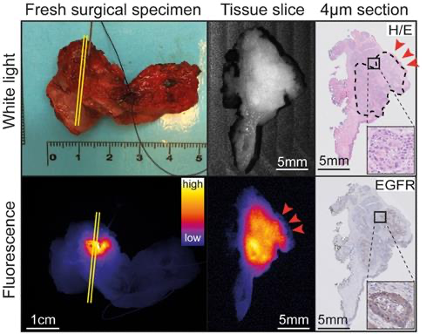
Molecular fluorescence-guided surgery may lead to increased detection of primary tumors, satellite lesions and lymph node metastasis, which allows clinicians to treat patients in earlier stages of disease. Moreover, this technique can be used intraoperatively by providing the surgeon with real-time feedback on the resection margin status, enabling radical excision of the tumor. Our expertise lies in early-phase clinical trials for the evaluation of novel tumor-specific fluorescent tracers for different applications. In fact, we are running one of the largest clinical trials to date in molecular fluorescence-guided surgery, focusing on ex vivo margin assessment during oral cancer surgery (NCT03134846). In all of our research we correlate our finding with histology.
Studies