Imaging techniques
Within the OMIG several state-of-the-art imaging instruments are available both for in vivo and ex vivo imaging. Also for preclinical applications in animal models, fluorescent probes and all imaging instrumentation (i.e. PET, SPECT, MRI, confocal laser endomicroscopy and optical systems) are available at the UMCG.
Intraoperative imaging systems
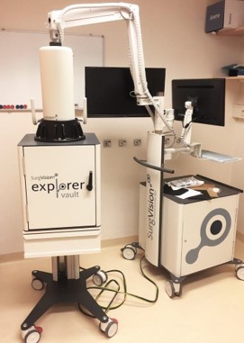
Clinical camera prototypes for intraoperative imaging have been developed, tested and approved by the Investigational Research Board within the consortium UMCG/TUM (Technical University of Munich, prof Ntziachristos). For example, a custom-build fiber-based NIR fluorescence endoscopy platform was developed with the University of Munich. This system provides real-time fluorescence and simultaneous white light imaging during endoscopy.
Other imaging techniques
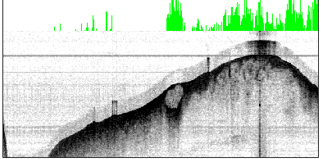
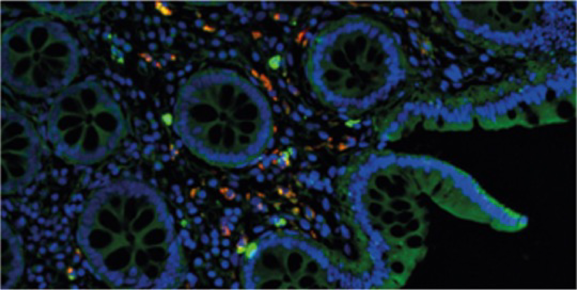
In addition, the fluorescence can be quantified in vivo using multi-diameter single fiber reflectance, single fiber fluorescence spectroscopy. For ex vivo tracer validation several systems such as the PEARL Imager or the Odyssey CLx flatbed scanner (both LI-COR) or fluorescence microscopy equipped for fluorescence in the NIR light spectrum are available. Besides the above mentioned imaging and quantification techniques we also use other novel techniques, such as optical coherence tomography and multispectral optoacoustic tomography.