Multispectral optoacoustic tomography
Multispectral optoacoustic tomography (MSOT) is an emerging real-time imaging technique that provides molecular information on the macroscopic and mesoscopic scale.
MSOT
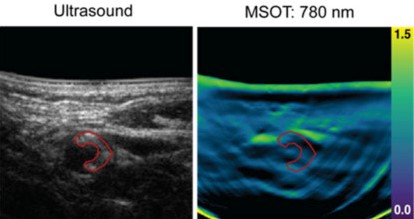
MSOT relies on the detection of ultrasound waves emitted by tissue due to absorption of light of transient energy. This contrast arises from both intrinsic tissue chromophores as well as exogenous contrast agents. Unlike fluorescence imaging, MSOT provides optical contrast to depths of several centimeters as the optoacoustic signal is less affected by scattering.
Ultrasound-MSOT in the OMIG
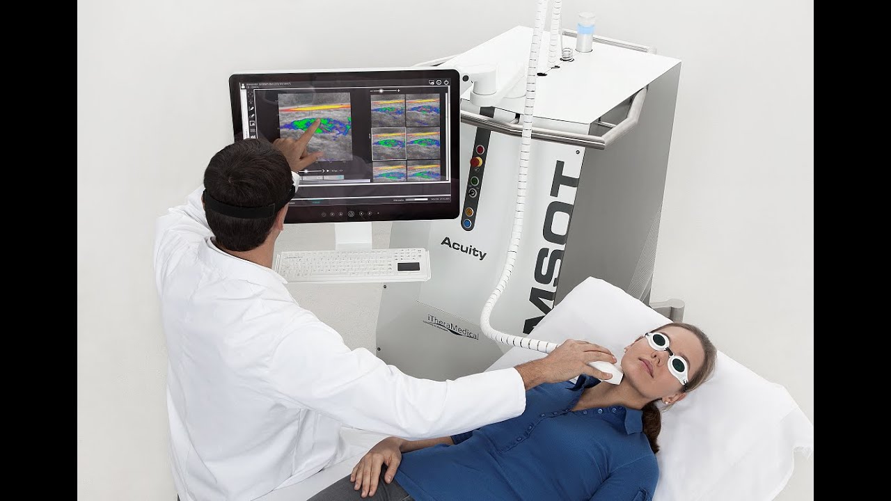
In the OMIG, we have a state-of-the-art clinical hybrid ultrasound-MSOT system (MSOT Acuity Echo, iThera Medical GmbH) available. Our main research focus is to explore new clinical indications for MSOT and perform functional and molecular imaging to enable earlier diagnosis and treatment stratification. Furthermore, we aim to develop optoacoustic tracers, which are currently not clinically available but have gained increasing interest.
Following data acquisition, the optoacoustic signal obtained needs to undergo reconstruction in order to obtain images. In our clinical MSOT studies, we closely collaborate with the Computation & Machine Learning research group at the Center for Translational Cancer Research Technical University of Munich (TranslaTUM). This group develops advanced data analysis tools for MSOT to extract accurate information and perform spectral analysis to separate different chromophores from the raw imaging data.