Optical Coherence Tomography
Optical coherence tomography (OCT) is a high-resolution optical imaging technique. By using this technique, we can capture micrometer-resolution images in 3D from within the tissue. In the ophthalmology, OCT is been used for a while and we would like to introduce the benefits of this technique in our field of research as well.
OCT in endoscopy
Optical coherence tomography (OCT) is the optical analog of ultrasound. It provides cross-sectional images of tissue by measuring the strength of backscattered light as a function of depth in tissue with a resolution of approximately 5-30 µm and a penetration depth of about 2-3 mm in tissue. Facilitating three-dimensional imaging at (sub)cellular level for millimeters into tissue, OCT is highly suited to the imaging of tissue epithelia. As a significant proportion of cancers form in the epithelia of hollow organs, the potential applications of OCT have a large scope. For instance, OCT may facilitate improvement in the detection rates of esophageal adenocarcinoma, for example, through the ability to detect both lesions and early indicators of dysplasia. Given the limited penetration depth of OCT, endoscopic use of the imaging technique must be applied in order to image in vivo.
OCT and fluorescence molecular imaging
While OCT provides high-resolution structural information in depth, it lacks in molecular contrast. To overcome this, we combined OCT with (Near Infrared) imaging together with the VU Amsterdam (prof. Dr. Johannes de Boer). In fact, the development of novel near-infrared fluorophores has opened the door to the possibility of fluorescently labeling a variety of monoclonal antibodies for targeted imaging.
The goal is to achieve molecular contrast combined with the structural OCT information to provide a paradigm shift in the kind of information available from minimally invasive imaging, approaching immunohistochemistry in vivo.
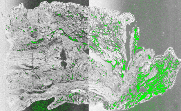
One of the first studies done with OCT by Prof. Dr. Johannes de Boer is described in this article: High resolution combined molecular and structural optical imaging of colorectal cancer in a xenograft mouse model (2018)