New publication by Madelon J. H. Metman, Pascal K. C. Jonker, Luc H. J. Sondorp et al in European Journal of Nuclear Medicine and Molecular Imaging

Our latest publication by Madelon J. H. Metman, Pascal K. C. Jonker, Luc H.J. Sondorp et al entitled ‘MET-receptor targeted fluorescent imaging and spectroscopy to detect multifocal papillary thyroid cancer’ can be found here.
Abstract
Purpose
Multifocal disease in PTC is associated with an increased recurrence rate. Multifocal disease (MD) is underdiagnosed with the current gold standard of pre-operative ultrasound staging. Here, we evaluate the use of EMI-137 targeted molecular fluorescence-guided imaging (MFGI) and spectroscopy as a tool for the intra-operative detection of uni- and multifocal papillary thyroid cancer (PTC) aiming to improve disease staging and treatment selection.
Methods
A phase-1 study (NCT03470259) with EMI-137 was conducted to evaluate the possibility of detecting PTC using MFGI and quantitative fiber-optic spectroscopy.
Results
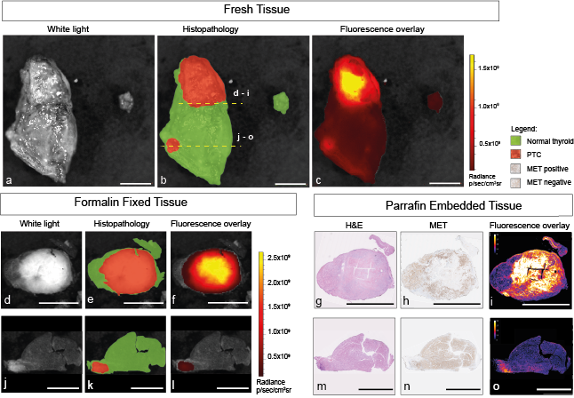
Fourteen patients underwent hemi- or total thyroidectomy (TTX) after administration of 0.09 mg/kg (n = 1), 0.13 mg/kg (n = 8), or 0.18 mg/kg (n = 5) EMI-137. Both MFGI and spectroscopy could differentiate PTC from healthy thyroid tissue after administration of EMI-137, which binds selectively to MET in PTC. 0.13 mg/kg was the lowest dosage EMI-137 that allowed for differentiation between PTC and healthy thyroid tissue. The smallest PTC focus detected by MFGI was 1.4 mm. MFGI restaged 80% of patients from unifocal to multifocal PTC compared to ultrasound.
Conclusion
EMI-137-guided MFGI and spectroscopy can be used to detect multifocal PTC. This may improve disease staging and treatment selection between hemi- and total thyroidectomy by better differentiation between unifocal and multifocal disease.