New publication by Van der Laan et al. in Cancers

Our latest publication by Jouke van der Laan entitled ‘Optical Biopsy of Dysplasia in Barrett’s Oesophagus Assisted by Artificial Intelligence’ can be found here.
Abstract
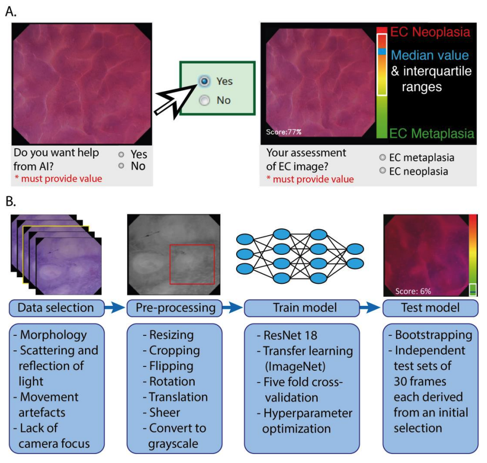
Optical biopsy in Barrett’s oesophagus (BE) using endocytoscopy (EC) could optimize endoscopic screening. However, the identification of dysplasia is challenging due to the complex interpretation of the highly detailed images. Therefore, we assessed whether using artificial intelligence (AI) as second assessor could help gastroenterologists in interpreting endocytoscopic BE images. First, we prospectively videotaped 52 BE patients with EC. Then we trained and tested the AI pm distinct datasets drawn from 83,277 frames, developed an endocytoscopic BE classification system, and designed online training and testing modules. We invited two successive cohorts for these online modules: 10 endoscopists to validate the classification system and 12 gastroenterologists to evaluate AI as second assessor by providing six of them with the option to request AI assistance. Training the endoscopists in the classification system established an improved sensitivity of 90.0% (+32.67%, p < 0.001) and an accuracy of 77.67% (+13.0%, p = 0.020) compared with the baseline. However, these values deteriorated at follow-up (−16.67%, p < 0.001 and -8.0%, p = 0.009). Contrastingly, AI-assisted gastroenterologists maintained high sensitivity and accuracy at follow-up, subsequently outperforming the unassisted gastroenterologists (+20.0%, p = 0.025 and +12.22%, p = 0.05). Thus, best diagnostic scores for the identification of dysplasia emerged through human–machine collaboration between trained gastroenterologists with AI as the second assessor. Therefore, AI could support clinical implementation of optical biopsies through EC.