New publication by Vonk, de Wit et al. in British Journal of Dermatology

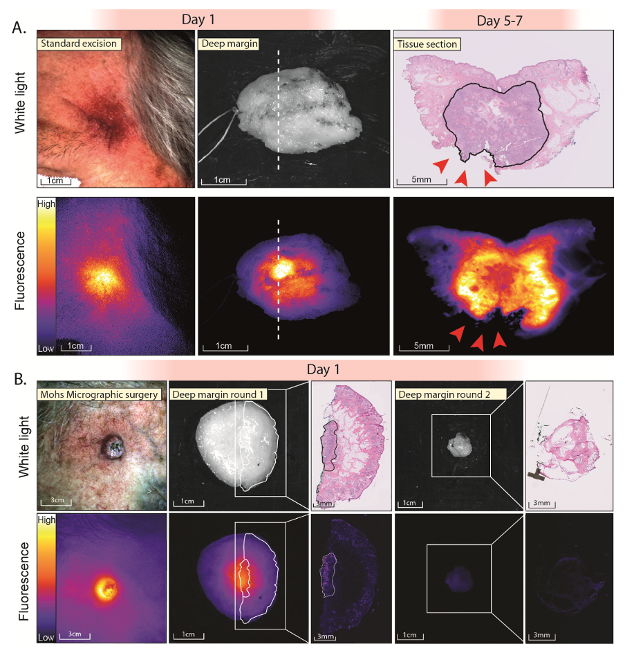
Our latest publication by Jasper Vonk and Jaron de Wit was published in the British Journal of Dermatology, showing the potential of fluorescence molecular imaging in the treatment of skin cancers. The full text can be found here.

Fluorescence imaging during standard excision and Mohs micrographic surgery
Figure 1: Fluorescence imaging during standard excision and Mohs micrographic surgery. A) Fluorescence imaging before standard excision of a temporal, s ub d e rma l tumo r that was earlier irradically removed. Ex vivo imaging of the specimen showed a fluorescent lesion at the deep resection ma rgin, c o rre la tin g with a tumo r-positive margin on histopatholo gy (red arrows). B) Fluorescence ima gin g during a MMS procedure. In vivo imaging shows a sharply demarcated fluorescent le s io n. Ex vivo imaging shows a fluorescent lesion at the deep resection margin, whic h co -lo c a lises with tumor on H&E histopatho lo gy. The second MMS round did not show any re ma inin g fluorescence signal, and no tumor was found on histopatho lo gy. Abbreviatio ns : H&E; Haematoxylin and Eosin, MMS; Mohs Micrograp h ic Surgery.